Discovery | Light-sheet
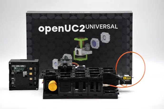
Das openUC2 LightSheet-Add-On ermöglicht den Aufbau eines Lichtblattmikroskops mit minimaler Komponentenanzahl und kompakter Stellfläche. Dieses Kit wurde für die nahtlose Zusammenarbeit mit der CoreBox, der ElectronicsBox, der FluoBox und der InfinityBox entwickelt und enthält alles, was für die automatisierte Lichtblatt-Bildgebung in biologischen und volumetrischen Anwendungen benötigt wird.
Enthaltene Komponenten:
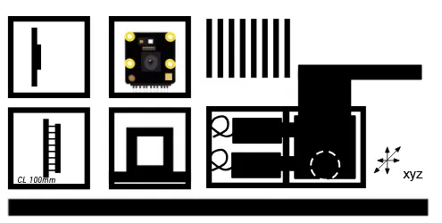
1x Zylinderlinse (100 mm, einstellbar zur Erzeugung des Lichtblatts)
1x Probenkammer (mit Wasser befüllbar, mit integrierter NeoPixel-LED zur Durchlichtbeleuchtung)
1x Mikrometer-XYZ-Tisch (präzise Probenpositionierung mit Spritzen-Probenhaltern)
1x abnehmbarer Emissionsfilter
1x zusätzliche Kamera (zur Unterstützung des Ausrichtungsprozesses)
1x massive Grundplatte (10 mm POM, 4×5 Würfel)
8x Puzzle-Grundplatten
Hinweis: Funktioniert nur in Kombination mit Discovery Core, Electronics-Add-On, Infinity-Add-On, FluoLaser-Add-On und Raspberry Pi mit Imswitch
openUC2 LightSheet Add-On
Does not work without CoreBox, Electronics Add-On, Infinity Add-On, Fluoresence Add-On Laser Edition and the Rasperry Pi Kit
Build a Fully Automated Light Sheet Microscope
The openUC2 LightSheet Add-On enables the construction of a light sheet microscope with a minimal component count and compact footprint. Designed to work seamlessly with the CoreBox, ElectronicsBox, FluoBox, and InfinityBox, this kit provides everything needed for automated light sheet imaging in biological and volumetric applications.
Included Components
In Cubes
- Cylindrical Lens (100mm, adjustable for creating the light sheet)
- Sample Chamber (water-fillable with integrated NeoPixel LED for transmitted light illumination)
- Micrometer XYZ Stage (precise sample positioning with syringe sample holders)
- removable Emission Filter
- additional Camera (to help with the allinging process)
Additional Accessories
- Solid Baseplate (10mm POM, 4x5 Cubes)
- 8x Puzzle Baseplates
Features and Applications
This kit is ideal for imaging biological samples, such as living organisms, cells, or tissues, in a 3D environment. Its modular design ensures easy integration into existing setups, allowing advanced light sheet microscopy with minimal adjustments.
- Compact Light Sheet Design: Based on the openSPIM configuration, this setup minimizes complexity while maintaining high performance. The entire assembly fits on a baseplate measuring only 25×20 cm.
- Automated Volume Imaging: Use the micrometer XYZ stage to move samples in all directions, enabling 3D imaging and volumetric scans.
- Light Sheet Generation: Collimate a single-mode, fiber-coupled 488nm laser and focus it into the back focal plane of a cylindrical lens. The astigmatism of the lens creates the light sheet, which illuminates the sample chamber.
- Orthogonal Fluorescence Detection: Perpendicular to the light sheet, the fluorescence imaging system uses an infinite-corrected objective, emission filter, tube lens, and monochrome camera to capture high-quality images.
- Software Integration: Controlled via the ImSwitch software and a streamlined UC2-eSP firmware, the system supports automated data acquisition and light sheet alignment.
Explore Light Sheet Imaging Today
With the openUC2 LightSheet Add-On, bring advanced light sheet microscopy to your lab with an open-source, fully customizable approach. Expand your microscopy horizons with ease and precision.
Three dimensional microscopy
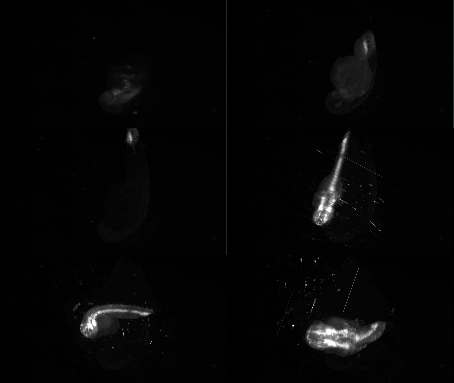
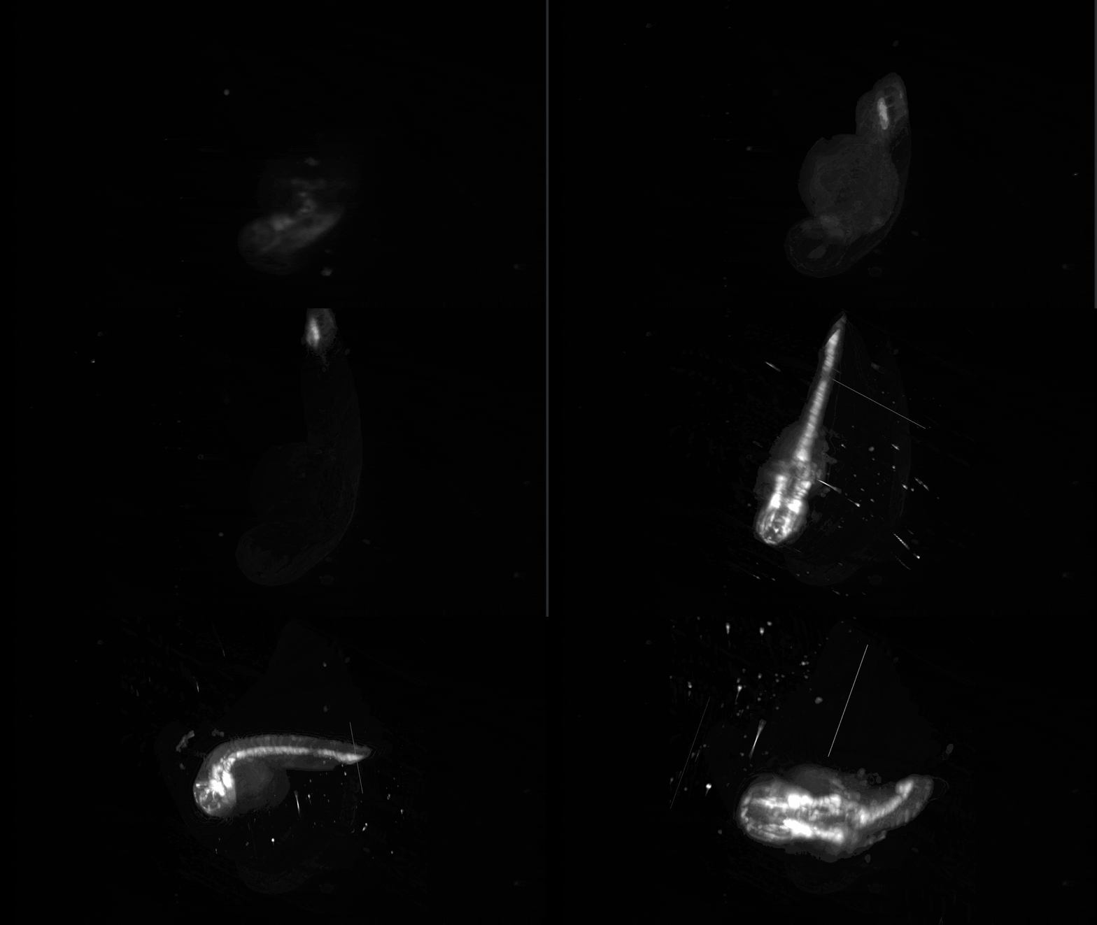
The light sheet technology enables fast, gentle, and high-contrast imaging of biological samples by illuminating only a thin slice of the specimen. By systematically shifting the sample in x, y, and z directions and stacking the resulting images, you can reconstruct detailed three-dimensional visualizations of microscopic structures.